
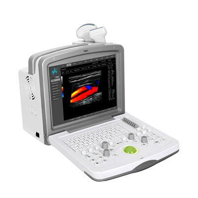
🩺 Core System Overview
Advanced fully‑digital Colour Doppler diagnostic ultrasound platform with high image quality and blood‑flow sensitivity.
Designed for multi‑specialty clinical applications including abdomen, OB/GYN, cardiology, small organs, superficial vessels, musculoskeletal, ophthalmology, anesthesiology, urology, and neurosurgery.
Portable, ergonomic design suitable for point‑of‑care use.
📊 Imaging & Performance
Imaging Modes:
B‑mode (2D imaging)
Colour Doppler
Triplex display (simultaneous B + Colour + Pulsed Doppler)
Elastography support (depending on configuration)
Advanced Imaging Technologies:
Pulse inversion harmonic imaging for improved contrast resolution
Sparse transmit & multi‑beam parallel processing for higher frame rates
Synthetic aperture beam forming
Speckle noise suppression to clean up 2D images
🧠 Hardware & Architecture
Processor: 8‑core DSP with latest generation front‑end ultrasound chip.
Display: Typically integrated medical LCD (e.g., 12″ size on most units).
Supports hot‑swappable probe connectors (usually 2 ports).
🔌 Connectivity & Networking
Multiple data transmission ports:
USB2.0, RS‑232, XGA
DICOM 3.0 for PACS connectivity and network transmission
DVD‑R/W (on some configurations)
🔎 Probes (Transducers)
Standard and optional probes cover a wide clinical range:
Standard: Convex (e.g., 5C2A, 2.0–5.0 MHz)
Optional:
Linear high‑frequency (e.g., 12L5A, 5.0–12.0 MHz)
Transvaginal (e.g., 8EC4A, 4.0–8.0 MHz)
Phased array (e.g., 4V2S, 2.0–4.0 MHz)
💾 Software & Workflow
Automatic parameter optimization based on exam type.
Versatile measurement software for abdominal, cardiac, OB/GYN, vascular, small organs, and musculoskeletal exams.
Supports export/storage of image formats (AVI, JPG, TIF, BMP, SEQ) and DICOM reports.
🖨️ User Interface
Ergonomic control panel with backlit controls and knobs.
Operates on a Windows‑based interface (familiar UI for workflow).
